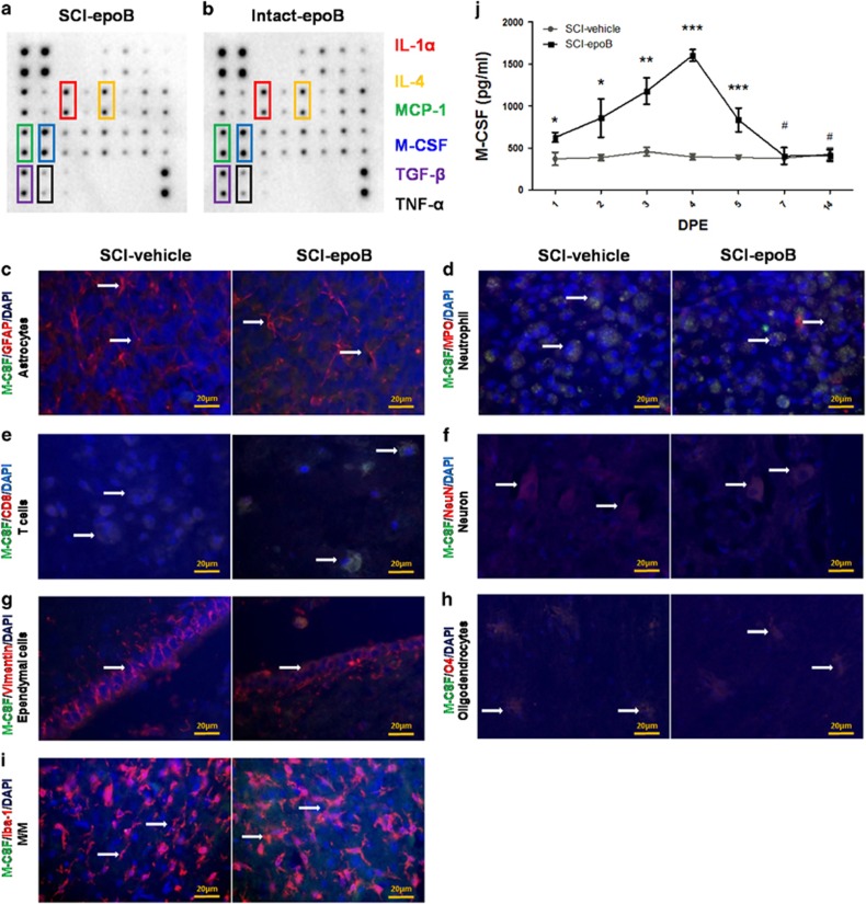
Figure 2.
Intact CNS astrocytes, T cells, ependymal cells, and M/Ms mainly contribute to the increasing secretion of M-CSF. (a and b) Two membranes show that the CSF cytokine profile of SCI-epoB (a) or intact-epoB (b) were unchanged in six cytokines 3 DPE. (c–i) Representative fluorescent staining images show SCI-epoB and SCI-vehicle 3 DPE. Double staining of particular cell types of CNS and M-CSF plus DAPI labeling for nuclei shows astrocytes (c), T cells (e), neurons (f), ependymal cells (g) and M/Ms (i) increase the secreting of M-CSF. Others cells of neutrophils (d) and oligodendrocytes (h) exhibit no change on M-CSF secretion. Arrows indicate examples of double-labeled cells. (j) Quantitative analysis of M-CSF expression in the CSF by ELISA. Means±S.D.; n=3 per group in (a) and (b). n=6 per group in (c)–(i). n=3 per group in (j); *P-value <0.05; **P-value <0.01; ***P-value <0.001 (one-way ANOVA)

