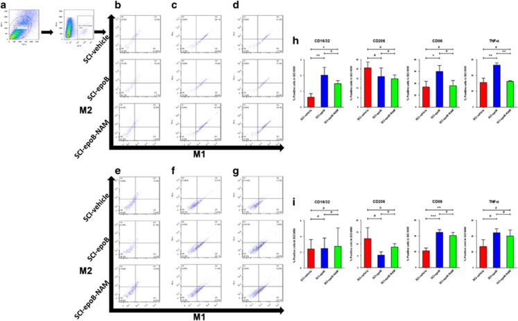
Figure 4.
SCI lesions polarize M/M from M2 to M1 phenotype 3 days after epoB treatment; the neutralizing antibody mix (NAM) partly reverses this polarization. (a) Representative plots showing the separating process of M/M in SCI lesions. Representative plots of M1 and M2 polarization are distinguished by (b) CD16/32 plus CD206, (c) CD86 plus CD163, and (d) TNF-α plus TGF-β in the SCI-vehicle, SCI-epoB, and SCI-epoB-NAM lesion sites 3 DPE. This experiment was conducted on the above group with the markers of (e) CD16/32 plus CD206, (f) CD86 plus CD163, and (g) TNF-α plus TGF-β 7 DPE. (h–i) Quantification of M/M polarization analyzed by CD16/32, CD86, CD163, and TNF-α at 3 DPE (h) and 7 DPE (i). Means±S.D.; n=6 per group; *P-value <0.05; **P-value <0.01; ***P-value <0.001 (one-way ANOVA)

