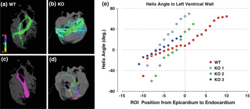
FIG. 6.

Comparison of myofiber helix angles in corresponding DSI projections in wild type (WT) compared with MyBP-C−/− (KO) hearts. Isolated subepicardial and subendocardial fibers of WT (red) and KO (blue, purple, green) hearts shown from the two chamber, left ventricle view. A ROI sphere was used to isolate fibers from DSI projections. (a and b) Display fibers on the subepicardial surface. (c and d) Display fibers on the subendocardial surface. While (a) and (b) both display coherent tracts, fibers on the subendocardium differ. The WT (c) fibers remain strongly coherent, while the KO (D) fibers are few and disorganized. Myofiber helix angles in the left ventricle of WT and KO hearts were imaged by DSI and plotted as a function of varying transmural locations in the case of individual WT and KO animals (e). The myofiber tracts in the WT myocardium varied from 260° helix angle in the subepicardium to 0° in the midmyocardium and 87° in the subendocardium. In the MyBP-C−/− myocardium, the transition from negative to positive helix angles occurs faster and more linearly than the wild type and is restricted to the subepicardium (negative ROI positions). From the midmyocardium to the subendocardium, the helix angles could not be measured because of a lack of coherence.
