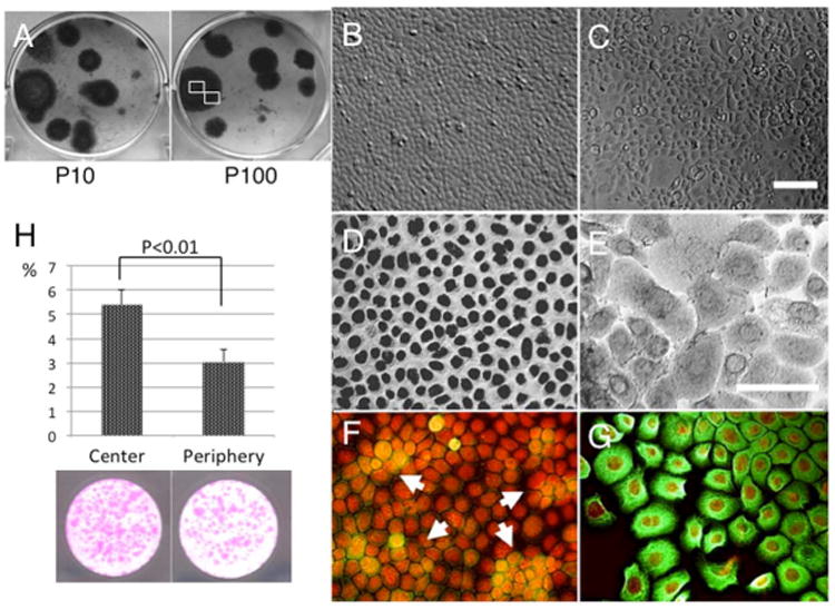
Figure 1. Progressive differentiation in the periphery of clonal growth.

Murine corneal/limbal epithelial progenitor cells exhibited clonal growth when seeded at 500 cells/cm2 in KSFM. Clones were round for both Passage 10 and 100 cultures (A = P10 and P100). In each P15 clone, cells in the center were uniformly small (A and B), but those in the periphery were enlarged and showed vacuolation (C). p63 nuclear staining was positive in all small cells in the center (D), but became weaker and also negative in larger cells in the periphery (E, marked by arrows). Expression of K14 keratin (green) was scanty in most small cells, but there was some sporadic staining of cells, particularly in the center (F, arrows). This was more uniformly positive in the cytoplasm of cells in the periphery (G). Bar represents 50 μm. Large-colony-forming efficiency (more than 0.5 mm diameter) was significantly higher in central location of the colony (5.38±0.6%) to comared with peripheral location (3.05±0.5%). (H, p<0.01)
