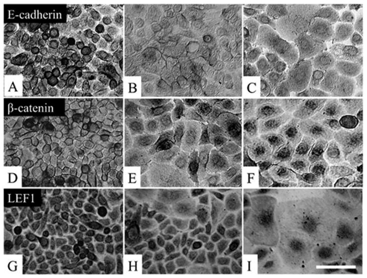
Figure 2. Activation of Wnt signaling during clonal expansion.

P15 clones were examined for activation of the Wnt signaling, Immunostaining to E-cadherin showed strong positive staining at the intercellular junctions by cells in the center (A). E-cadherin expression became more in the cytoplasm for some cells in the mid-periphery (B), and it was located exclusively in the perinuclear cytoplasm in the periphery of the clone (C). β-catenin was also located intercellularly in the center (D), but it became located in the perinuclear cytoplasm in the mid-periphery (E), and it was exclusively in the nucleus in the periphery (F). LEF-1 was in the cytoplasm in cells in the center (G), but staining became nuclear for some cells in the mid-periphery (H), and for all cells in the periphery (I). Bar represents 50 μm.
