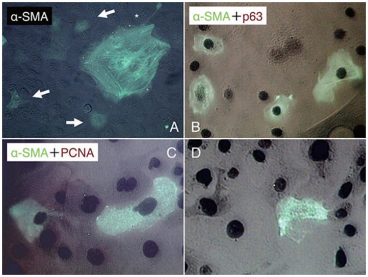
Figure 4. Loss of proliferative activity during EMT.

In P15 clones, α-SMA expression was observed in association with cytoplastic stress fibers in large squamous cells (A, marked by *), and also, it was found in the cytoplasm in some small to intermediate cells without stress fibers (A, marked by arrows). Double immunostaining to α-SMA (green) and p63 (brown) showed that some of the α-SMA-expressing small/intermediate cells still expressed nuclear p63 (B). Double immunostaining to α-SMA (green) and PCNA (brown) also showed that α-SMA-positive cells in the mid-periphery also had a positive nuclear staining for PCNA (C). Cells in periphery lost their nuclear staining for PCNA (D). Bar represents 50 μm.
