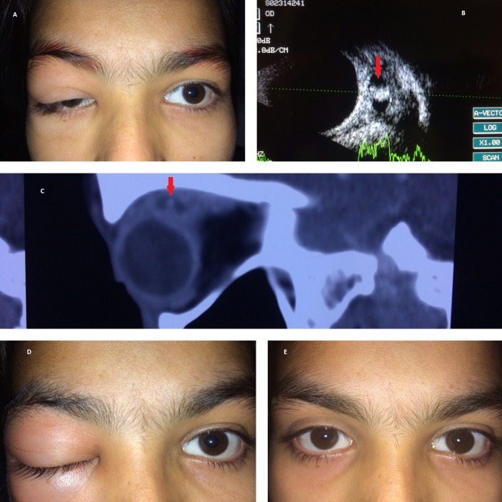
Figure 1.
(A) Initial clinical presentation with recurrent swelling and drooping of upper eyelid. (B) Ultrasonography of the superior rectus muscle showing a large cyst with a central high-amplitude spike corresponding to the scolex (red arrow). (C) Sagittal view of the non-contrast CT of the orbit showing inflammatory thickening of the superior rectus muscle with the cystic cavity having the central scolex (red arrow). (D) Inflammatory swelling of the right periorbital region after the patient skipped oral steroids. (E) Complete recovery of ptosis and inflammatory signs at the end of 4 weeks.

