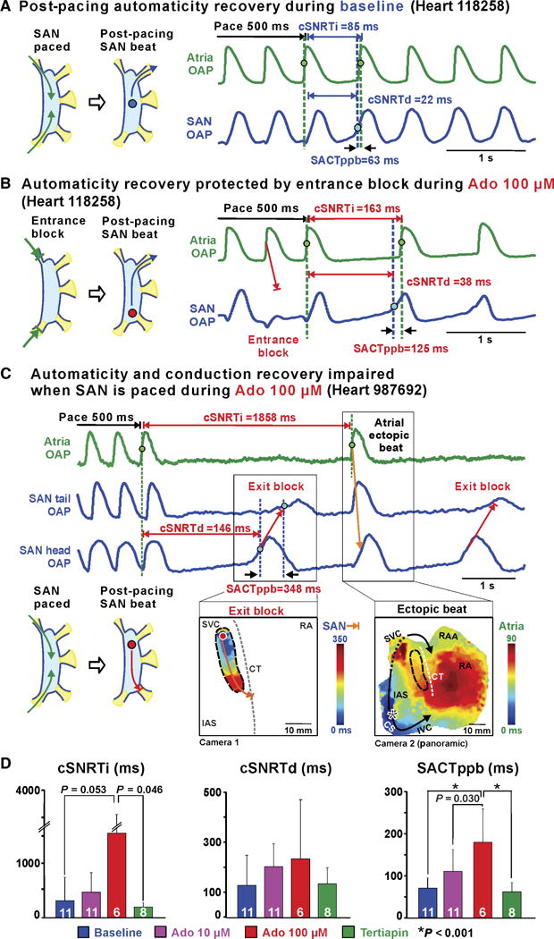
Fig. 5. SAN intranodal automaticity and conduction strongly affected by combination of atrial pacing and adenosine in ex vivo human hearts. (A).

Baseline SAN activation and conduction pattern during and after atrial pacing. (B) Adenosine impaired SAN automaticity and conduction; conversely, entrance block, caused by conduction depression, prevented overdrive suppression of SAN automaticity. (C) Top: Atrial and SAN OAPs during 500-ms CL pacing and post-pacing with 100 μM adenosine. Bottom: SAN exit block (left) and atrial ectopic beat (right) activation maps during 100 μM adenosine. (D) Bar graphs summarizing average drug effect on cSNRTi (left), cSNRTd (middle), and SACTppb (right). White numbers show the number of hearts used. Data are means ± SD.P values were determined by ANOVA after Tukey’s adjustment. CS, coronary sinus; RAA, right atrial appendage; SACTppb, sinoatrial conduction time of first post-pacing SAN beat; SNRTi/d, indirect/direct SAN recovery time; cSNRTi/d, corrected SNRTi/d.
