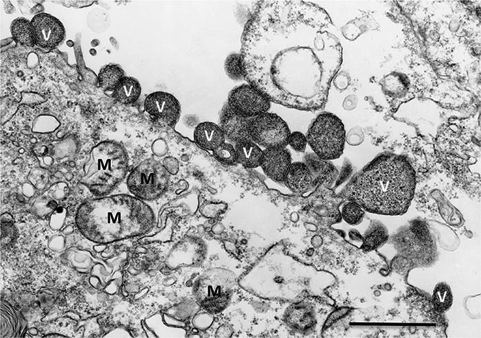
Fig. 1.

Transmission electron micrograph of Vero E6 cells infected with Sierra Nevada virus. High magnification of virions (V) budding from the cell surface. Mitochondria (M) are indicated for reference. Scale bar=1 µm. (Contributed by Dr Vsevolod Popov, Department of Pathology, Center for Biodefense and Emerging Infectious Diseases, University of Texas Medical Branch, Galveston, TX, USA).
