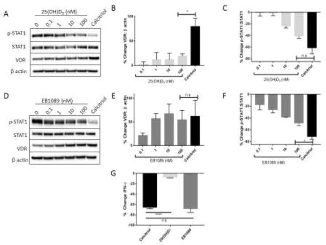
Figure 4. VDR upregulation is necessary for reduction in IFN-γ but not p-STAT1 after treatment with vitamin D or analogs.
TL-1 cells were treated with increasing dosages of the inactive form of vitamin D, 25(OH)D3 (A–C), or the high affinity calcitriol analog EB1089 (D–F), as well as vehicle control or calcitriol for comparison. A, D. Protein lysates were prepared and western blot analysis was performed (n=3, +/− SEM). Representative western blots are shown. Protein levels were normalized to β actin for VDR (B, E) or STAT1 for p-STAT1 (C, F), and then further normalized to the vehicle control to illustrate changes as a result of calcitriol treatment. G. Conditioned media was collected from the 100 nM calcitriol, 25(OH)D3, and EB1089 samples after 24 h and submitted for Luminex cytokine analysis (n.s=not significant, *p<0.05, ****p<0.001).

