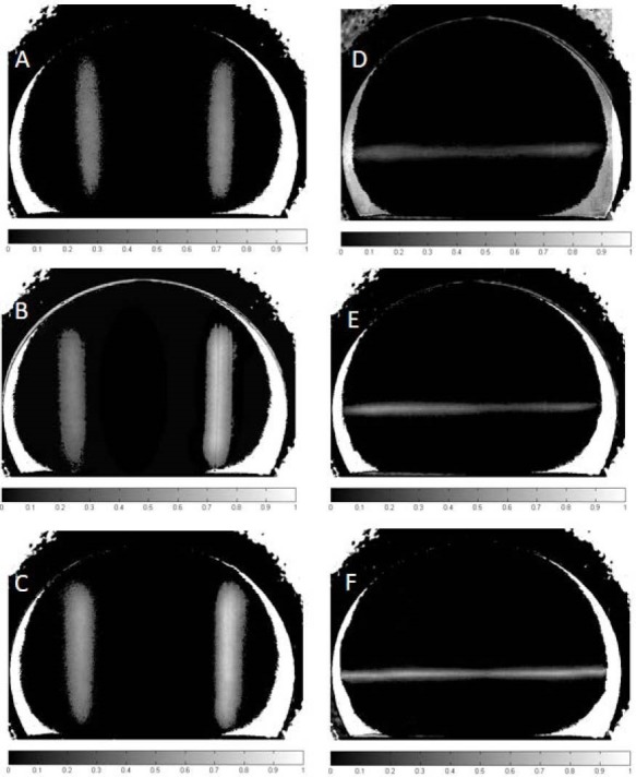
Figure 3.

Processed Images of Phantom for Major and Minor Vessels. (A) Image of phantom when left and right major vessels were filled by 1× and 2× concentration of Hb; (B) 1× and 4× concentration of Hb; (C) 2× and 4× concentration of Hb; (D) Image of phantom when minor vessels were filled by 1× Hb; (E) 2× concentration of Hb; (F): 4× concentration of Hb.
