Fig. 2.
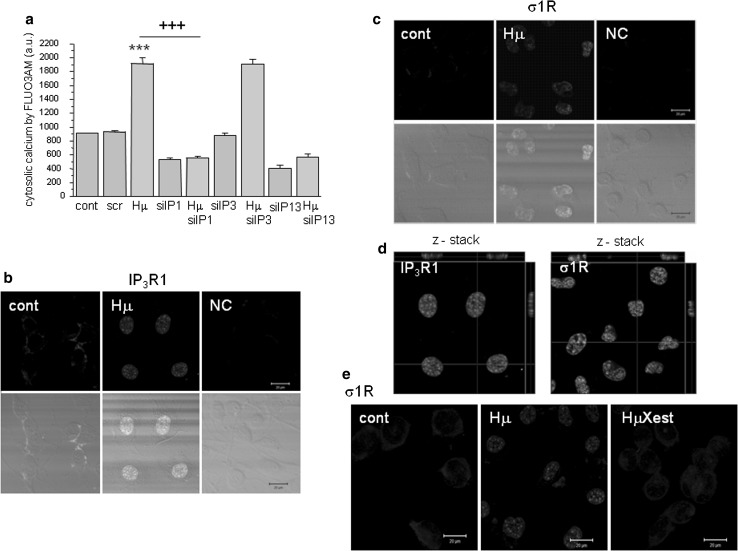
Involvement of IP3R1, but not IP3R3 in haloperidol-induced changes in levels of cytosolic calcium and translocation of the IP3R1 and σ1R to the nucleus following haloperidol treatment. Experiments were performed on NG-108 cells differentiated by dbcAMP. To determined haloperidol-induced changes in cytosolic calcium due to IP3R1/IP3R3 receptors, these receptors were silenced either individually, or both of them and levels of cytosolic calcium levels were determined after haloperidol treatment (a). Silencing of IP3R1, but not IP3R3 caused significant decrease in cytosolic calcium levels, thus proving involvement of the IP3R1, but not IP3R3 in haloperidol-induced increase in cytosolic calcium. In control cells (cont), IP3R1s (b, green signal) and σ1Rs (c, green signal) are localized to the ER. Following haloperidol treatment (Hμ; 10 µmol/L), these receptors translocate to the nucleus (b, c, green signal). Translocation of the IP3R1 and σ1R was verified by z-stacks from the Hμ-treated cells (d), which clearly shows an intranuclear signal. In the presence of Xestospongin C (Xest; 1 µmol/L), the Hμ -induced translocation of σ1R does not occur (e, HμXest). Results in the graph are expressed as the mean ± SEM and represent an average of six parallels from two independent cultivations. Statistical significance compared to control was ***p < 0.0001 and compared to Hμ treated cells was +++p < 0.0001

