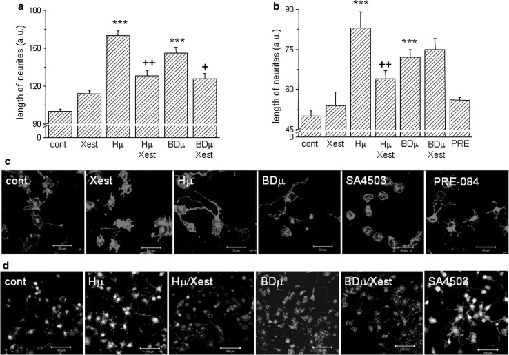
Fig. 6.
Effect of haloperidol (Hμ) and BD1047 (BDμ) on the length of neurites. Experiments were performed on NG-108 cells differentiated by dbcAMP. The cells were treated for 24 h with Hμ (10 µmol/L), BDμ (10 µmol/L) and Xest (1 µmol/L) after 72 h of differentiation. Length of neurites was measured either manually by ImageJ program (a), or using “Neurite outgrowth staining kit” (b–d). By both methods it is clearly shown that Hμ increased the length of neurites and this increase is partially prevented by Xest. Similar increase in length of neurites was visible after the BDµ treatment, although the effect of Xest was not so conclusive (a, b). Results from the confocal microscopy without a cell viability staining (c; bar represents 50 µm) or together with the cell viability stain (d; bar represents 100 µm) supported results from the fluorescent reader. Each column represents an average of 450–835 cells, and the results are displayed as the mean ± S.E.M. Statistical significance compared to controls is *p < 0.05, **p < 0.01, and ***p < 0.001 vs. control and +p < 0.05; ++p < 0.01, and +++p < 0.001 vs. Hμ or BDμ-treated cells

