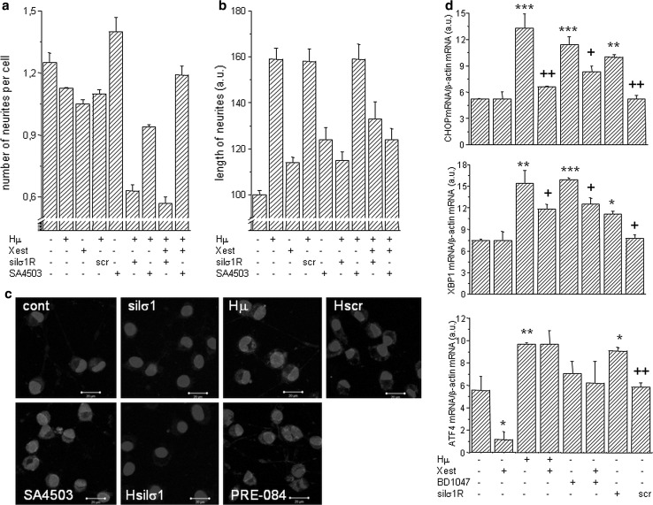
Fig. 7.
The impact of haloperidol treatment on cell’s plasticity. Experiments were performed on NG-108 cells differentiated by dbcAMP. The pharmacological impact was determined by measuring the number of neurites per cell (a) and neurite outgrowth (b) in haloperidol (Hμ; 10 µmol/L)-treated cells with a parallel treatment of Xestospongin C (Xest; 1 µmol/L), SA4503 (1 µmol/L) and with silenced σ1R. Number of neurites decreased significantly in Hμ-treated cells with silenced σ1R (a), but not with a scrambled siRNA. Silencing of the σ1R in Hμ-treated cells decreased significantly compared to plain Hμ-treated cell, similarly as by σ1R agonist SA4503 (b). Effectivity of σ1R silencing was verified by immunofluorescent staining (c). Bar represents 20 µm. Induction of markers of ER stress in Hμ, BDμ, and siRNA σ1R -treated differentiated NG-108 cells (d). The relative mRNA levels of CHOP, XBP1, and ATF4 were determined in control (cont), Hμ-treated and BDμ cells with or without Xest, then in cells after silencing of the σ1R (silσ1R) and scrambled siRNA (scr). A significant increase compared to control was observed in silσ1R cells and in Hμ- and BDμ-treated cells, but not in combination of these compounds with Xest. The results are expressed as the mean ± SEM. Statistical significance: * p < 0.05, ** p < 0.01, and *** p < 0.001 compared to untreated control cells; + p < 0.05, ++ p < 0.01 compared to Hμ, BD, and/or silσ1R treated group

