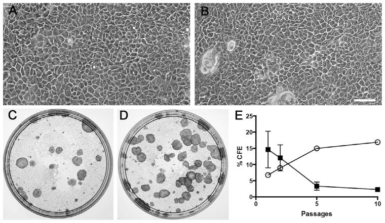
FIGURE 3.
Increased long-term proliferative potential in keratinocytes from Van der Woude syndrome (VWS) when compared with nonsyndromic cleft lip and palate (NSCLP). A, B: Phase contrast micrographs of keratinocytes obtained from the skin of children with NSCLP (A) or VWS (B). C to E: Macrographs of colony forming efficiency (CFE) assay performed with keratinocytes from children with NSCLP (C) or VWS (D). E: Average of CFE over 10 culture passages (solid square, NSCLP, n=7 at passage 1 [P1], n=6 at P2, n=5 at P5, n=1 at P10; open circle, VWS, n=1). Data are means ± SEM. Scale bar = 100 μm.

