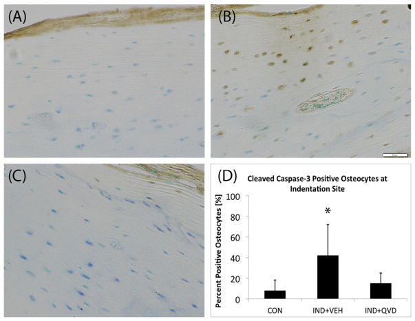
Fig. 5.
Role of osteocyte apoptosis in response to in vivo RPI loading. Representative IHC stained cross-sections of paraffin embedded mouse tibia, at 20× magnification showing (A, C) the absence of positively stained osteocytes in CON and IND + QVD groups and (B) clear evidence of positively stained osteocytes, morphological evidence of co-located intracortical resorption activity, in the IND + VEH group. (D) Shows data of percentage of positively stained osteocytes in samples from all three groups.

