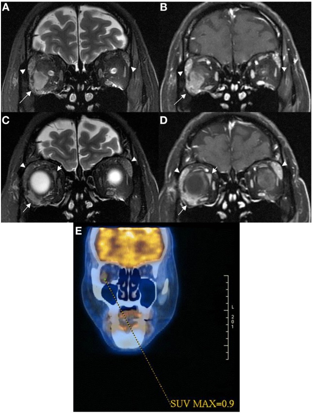
Figure 2.

Coronal MRI of patient 2 showed intraconal orbital mass lesion (arrow) with low T2W signal (A) and modest contrast enhancement (B) having a 0.9 SUV on 18FDG PET image (E). Coronal MRI more anteriorly showed extraconal orbital lesions (short and long arrows) with low T2W signals (C) with modest contrast enhancement (D). One of the extraconal lesion infiltrated the outer lateral part of the right globe [long arrows in (C,D)]. Lacrimal glands were enlarged [arrowheads in (A–D)], and they demonstrated low T2W signals with modest contrast enhancement suggestive of lymphoid tissue infiltrate.
