
Figure 1.
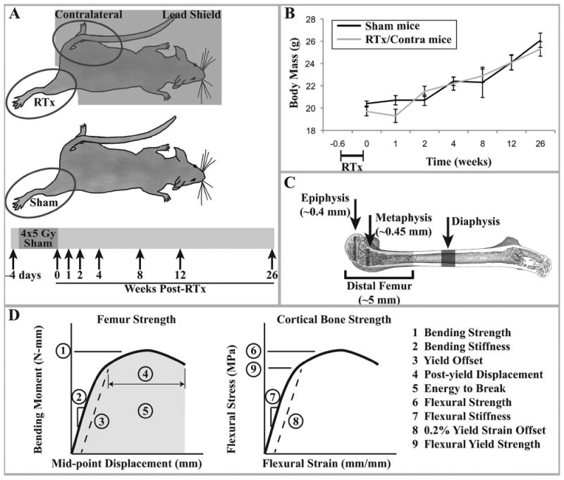
A) Experimental design: female BALB/cJ mice aged 12 weeks were exposed to four consecutive daily unilateral hindlimb irradiation exposures of 5 Gy each. The contralateral hindlimb and body were shielded with lead. These animals yielded the RTx and Contra samples. A separate group of age-matched animals were anesthetized but not irradiated, yielding the Sham samples. At end points of 4 days prior to treatment and 0, 1, 2, 4, 8, 12, and 26 weeks after treatment, animals were euthanized and femurs collected. B) Average mouse body mass for each treatment group (arithmetic mean ± standard error) over the course of the study. There were no significant differences in body mass at any time point between the sham and irradiated mice (contralateral femurs are derived from the non-irradiated limb of RTx group mice). C) Schematic of the μCT volumes of interest. D) Graphical description of the femur strength and cortical bone strength outcome measures.