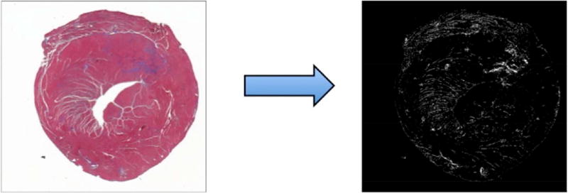
Figure 1.
Histology tissue section with trichrome stain (left) and post-processed histology image (right) are shown. Matlab color thresholding tools allow calculation of total myocardial pixels as the sum of non-white (e.g. red-myocardium and blue-collagen) pixels on the original image (left). Blue-staining collagen is rendered as white pixels on the thresholded image (right). Collagen volume fraction (CVF) is computed ratio of collagen-stained pixels to the total pixels.

