
Fig. 15.
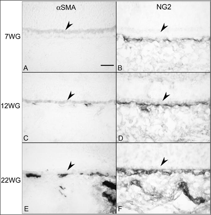
Alpha smooth muscle actin (αSMA) (A, C & E) and NG2 labeling (B, D & F) in choroid at 7 (A,B), 12 (C,D) and 22 WG (E,F). No immunoreactivity for αSMA (A) was observed at 7 WG but NG2 (B) labeled cells were associated with the choriocapillaris. Some scattered αSMA positive cells (C) were associated with choriocapillaris at 12 WG and NG2 staining (D) was more intense. αSMA immunoreactivity (E) was associated with choriocapillaris at 22 WG and was present in medium and large choroidal vessels. NG2 labeling was intense in all vessels. (Scale bar = 30 μm) (Fig. 6 from Baba et al., Invest. Ophthalmol. Vis. Sci. 50:3507, 2009, with permission).