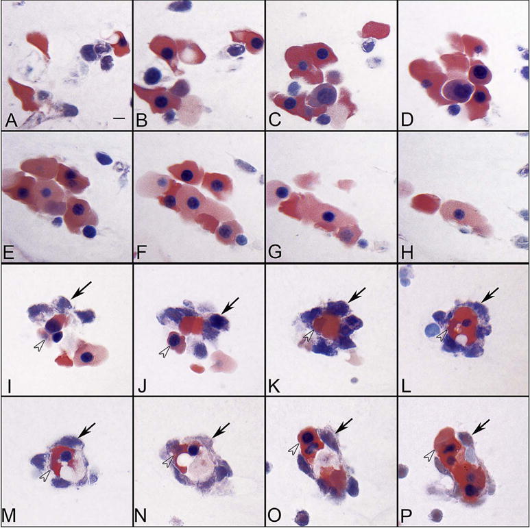
Fig. 2.

Blood island-like structures in serial sections of the vitreous at 5.5 WG. Some island-like structures are aggregates of erythroblasts (A–H), while others were composed of basophilic mesenchyme (arrows) (I–P) around a core of acidophilic erythroblasts (arrowheads). (Wrights-Giemsa stain, Scale bar in A = 10 μm for all) (Fig. 2 from McLeod et al., Invest. Ophthalmol. Vis. Sci. 53:7915, 2012, with permission).
