
Fig. 26.
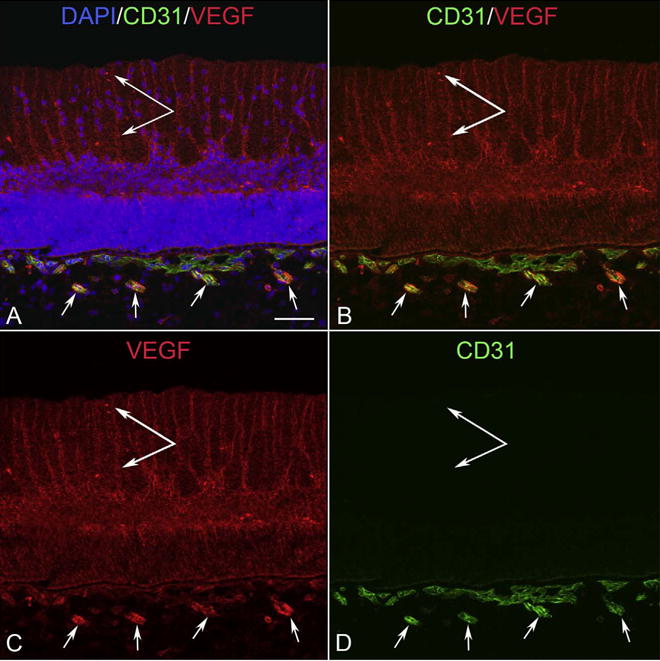
VEGF localization at 12 WG. VEGF was prominently localized to apparent Muller inner processes (double arrow), the inner neuroblastic layer in retina, and in intermediate blood vessels in Sattler’s layer of choroid that are developing at this time. There is less VEGF in primitive choriocapillaris. (Scale bar = 50 μm).