
Fig. 3.
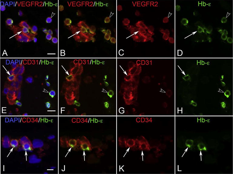
VEGFR2 (A–D), CD31 (E–H) and CD34 (I–L) labeling (all red) with epsilon hemoglobin (green, Hb-ε) and DAPI (blue, nuclei) in blood island-like structures in the 6 WG vitreous. Free erythroblasts co-expressed VEGFR2 and Hb-ε (arrowhead in A–D) as did some cells within blood islands (arrow in A–D). All cells of the blood islands expressed VEGFR2. CD31 and Hb-ε were co-expressed in select cells of blood islands (arrow in E–H) while free Hb-ε+ erythroblasts were only weakly immunoreactive for CD31 (arrowhead in E–H). CD34 and Hb-ε were co-expressed in some scattered cells in blood island-like structures (arrows in I–L), while free Hb-ε+ erythroblasts were CD34− (not shown). (Scale bar in A, E & I = 10 μm) (Fig. 5 from McLeod et al., Invest. Ophthalmol. Vis. Sci. 53:7918, 2012, with permission).