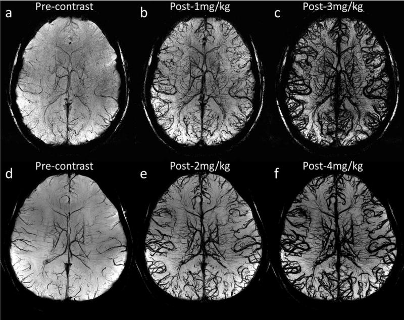
FIGURE 3.
Visualization of arteries and veins using minimum intensity projections of SWI with different doses of ferumoxytol. The effective slice thickness is 20 mm for all images. a to c were reconstructed using Volunteer 3’s data with voxel size 0.13 × 0.26 × 0.8 mm3, while d to f were reconstructed using Volunteer 1’s data with voxel size 0.11 × 0.22 × 1.25 mm3. See Table 1 for other imaging parameters for these two volunteers’ data.

