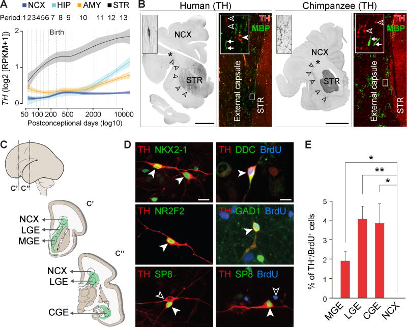
Fig. 5. Human telencephalic TH+ interneurons are of subpallial origin and start to express TH protein perinatally.
(A) TH expression in human neocortex (NCX), HIP, AMY, and STR throughout development. The shaded area corresponds to a confidence interval of 50%. (B) Immunohistochemistry reveals TH+ axons in external capsule (arrowheads), STR, and NCX of newborn (38 pcw) human and chimpanzee brains. Bipolar TH+ interneurons (filled arrowhead) are present in parallel with MBP+/TH− (arrows) and TH+/MBP− (open arrowheads) fibers in the external capsule. No TH+ cells were detected in chimpanzee external capsule. Scale bar represents 1 cm. (C) Schematic of dissection of ganglionic eminences (lateral [LGE], medial [MGE], and caudal [CGE]) and neocortical proliferative zones (NCX) from mid-fetal brain for primary cell culture. (D) TH+ cells from ganglionic eminences also express NKX2-1, NR2F2, or SP8, and were BrdU+, DDC+, and GAD1+. TH+ interneurons in the neocortical culture were SP8+, but BrdU− (bottom right panel). Scale bar represents 20 µm. (E) Percentage of TH+/BrdU+ cells in culture from MGE, LGE, CGE, and NCX. Error bars represent SEM. Pairwise t-tests were performed and corrected for multiple testing using Bonferroni correction. * P < 0.05; ** P < 0.01.

