
Figure 2.
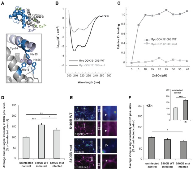
S100B sequesters zinc ions in hippocampal cells in vitro. (A) Illustration of the structure of S100B (PDB code 3d0y) showing the α-fold and zinc (purple sphere) and calcium (green spheres) binding sites; the enlargement represents the structural positions of residues involved in zinc interactions that were mutated to serines in the S100B mut variant. (B) Far-UV Circular Dichroism (CD) spectra of purified Myc-DDK-tagged wt S100B and mutant S100B at 0.1 mg.mL−1 in 50 mM TRIS-HCl pH 7.4, with typical α-helix minima observable at 208 and 222 nm. (C) Relative zinc binding to Myc-DDK tagged wt S100B (dark gray squares) is abolished in the Myc-DDK tagged S100B mutant (light gray squares), as inferred from PAR titration assays of S100B proteins (10 μM) with increasing ZnSO4 (at up to 40 μM) concentration. (D) Hippocampal neurons were infected at DIV1 with Lenti virus mediating expression of DDK-S100B wt, or DDK-S100B mut. The mean signal intensity of Zinquin fluorescence normalized to the co-localizing S100B fluorescence intensity is shown at DIV14. S100B is visualized by its anti-DDK tag. Cells expressing the mutant protein show significantly lower Zinquin signals at DDK-S100B mut positive sites compared to DDK-S100B WT positive puncta (one-way ANOVA, F = 19.347; p < 0.001; Post hoc analysis: control vs. S100B wt, p < 0.001; control vs. S100B mut, p = 0.004; S100B wt vs. S100B mut, p = 0.032). (E) Exemplary images showing infected cells. Zinquin signals are shown color-coded. Fluorescence intensities are assigned RGB colors. In S100B wt expressing neurons, a co-localizing Zinquin signal can be seen for S100B (upper panel). In cells expressing the mutated S100B, the fluorescence intensity of the Zinquin signal is significantly reduced (lower panel). S100B was visualized by anti-DDK antibody labeling (shown in magenta). (F) Addition of ZnCl2 has no effect on Zinquin signals associated with wild type S100B, and also does not lead to an increase in Zinquin signal associated with mutated S100B. Infected hippocampal cultures were treated with 60 μM ZnCl2 for 1 h. Addition of zinc significantly increases Zinquin fluorescence in control cells (insert; t-test, p < 0.0001). No further increase in Zinquin signal intensity was seen for wt S100B or mut S100B that showed significantly lower co-localizing Zinquin signal intensity (n = 10 cells; one-way ANOVA, F = 3.988; p = 0.03; Post hoc analysis: control vs. S100B mut, p = 0.024).