
Figure 3.
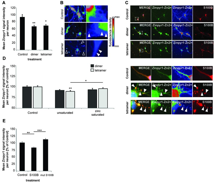
Increased levels of S100B sequester zinc ions. (A) Treatment of hippocampal cultures with 30 μM dimeric and with 30 μM tetrameric S100B for 24 h at DIV10 significantly reduces the intracellular Zn2+ concentration assessed by the analysis of Zinpyr1 signal intensity levels from 20 cells per condition for both application of the dimer and tetramer (Welch’s ANOVA, F = 8.084; p = 0.001; Post hoc analysis: control vs. dimer, p = 0.008; control vs. tetramer, p = 0.015). (B) Exemplary images showing signal intensities of Zinpyr1 in color—coded manner. Note the occurrence of Zn2+ containing clusters within neurons in cells treated with dimer and tetramer (full arrow) and extracellular Zn2+ positive accumulations (open arrow). (C) S100B signals co-localize with Zinpyr1 signals (full arrow) associated with Zn2+ after application of 30 μM dimeric or 30 μM tetrameric S100B for 24 h at DIV10. (D) Pre-incubation of S100B with 60 μM zinc leads to partial saturation of S100B zinc binding before application of 30 μM dimeric and with 30 μM tetrameric S100B for 24 h at DIV10, and thus to a significantly less decrease of intracellular zinc levels after treatment with S100B (Dimer: one-way ANOVA, F(2,45) = 2.611; p = 0.085; Tetramer: F(2,45) = 5.173; p = 0.01; Post hoc analysis: control vs. tetramer, p = 0.0045; tetramer vs. zinc saturated tetramer, p = 0.036; n = 16 cells). (E) Treatment of hippocampal cultures with 30 μM Myc-DDK-S100B wt, or Myc-DDK-S100B mut for 24 h at DIV10 leads to a significant decrease in intracellular Zinpyr1 fluorescence for wt S100B but not for the mutant S100B (one-way ANOVA, F(2,77) = 8.86; p < 0.0001; Post hoc analysis: control vs. S100B, p = 0.0037; control vs. S100Bmut, p = 0.2469; S100B vs. S100Bmut, p < 0.0001).