Figure 1.
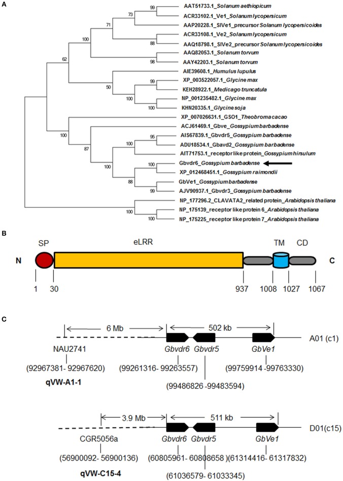
Phylogenetic and structural analysis of Gbvdr6 gene. (A) Phylogenetic relationship of Gbvdr6 with other Ve-like proteins by MEGA6 software with the neighbor-joining (NJ) algorithm under 1,000 replicates of bootstrap. The numbers on the internal nodes are the percentage bootstrap support values. (B) Schematic diagram of Gbvdr6 protein domain architecture showing signal peptide (SP) at N-terminus, followed by extracellular leucine-rich repeat (eLRRs), transmembrane (TM) domain and cytoplasmic domain (CD) at C-terminus. The numbers indicate the domain regions. (C) Schematic diagram of physical locations of Gbvdr6, Gbvdr5, and GbVe1 and the SSR markers flanking the known Verticillium wilt-resistant QTLs in the chromosomes of tetraploid cotton. The numbers in brackets indicate the physical positions in the chromosome. NAU2741 and CGR5056a are the SSR markers that flank Verticillium wilt resistance QTLs qVW-A1-1 and qVW-C15-4 in the A01(c1) and D01(c15) chromosomes, respectively.

