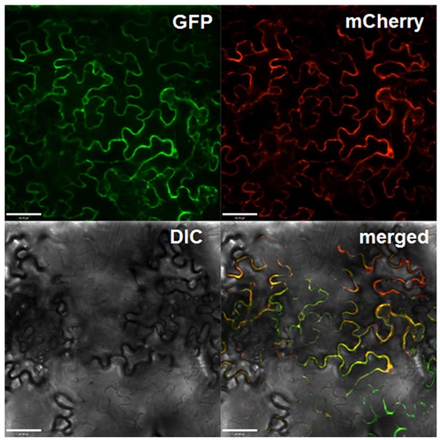
Figure 3.

Subcellular localization of Gbvdr6 in epidermal cells of N. tabacum leaves. The Gbvdr6-GFP fusion was transiently co-expressed with the plasma membrane marker mCherry. The images were taken under a confocal microscope at 48 h after agro-infiltration. GFP: fluorescence of Gbvdr6-GFP fusion, mCherry: fluorescence of the plasma membrane marker mCherry, DIC, differential interference contrast; merged, a merged image. Scale bar = 60 μm.
