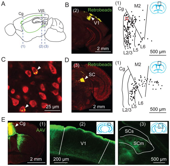
Fig. 1. Cg projects to visual cortex and superior colliculus (SC).
(A) Schematic of Cg projections. Dashed lines, locations of coronal sections shown in this figure: (1), Cg; (2), V1; (3), SC. (B to D) Retrograde tracing. (B) Left, Fluorescence image at location (2) showing Retrobeads (green) injected into V1. Arrowhead, injection site. Red, Nissl staining. Right, labeled neurons (dots) at (1), in region outlined by black rectangle (inset). (C) Fluorescence image for red square in (B). Arrowheads, labeled neurons. (D) Similar to (B), with Retrobeads injected into SC. (E) Anterograde tracing from Cg. Left, Fluorescence image at (1). Arrowhead, AAV injection site; middle and right, Cg projections to V1 and SC. SCs/SCm, sensory/motor related SC.

