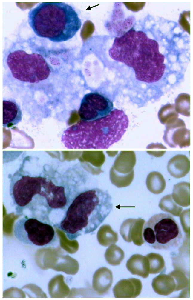
Figure 1.

Hemophagocytosis in the bone marrow during Leuconostoc pseudomesenteroides-associated hemophagocytic lymphohistiocytosis. Hypocellularity and active hemophagocytosis (arrow in the upper and lower panel) were identified in bone marrow aspirate. Magnification, ×1,000. Wright-Giemsa stain indicated phagocytosis of nucleated red blood cells and platelets. Cytoplasm of basophilic erythroblasts were indicated blue and nuclei of basophilic erythroblast were indicated purple.
