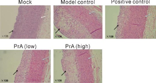
Figure 1.

Histological analysis by hematoxylin/eosin staining. Rabbits were then randomly divided into five groups (n=12): Mock, rabbits were offered continued normal diet; Model control, rabbits were offered continued high fat diet; Positive control, rabbits were fed with high fat diet containing 0.5 mg/kg rosuvastatin once every day; PrA low, rabbits were fed with high fat diet containing 5 mg/kg PrA once every day; PrA high, rabbits were fed with high fat diet containing 25 mg/kg PrA once every day. After 42 days, large or medium arteries were collected and subjected to HE staining. The dark arrow represents end arterium, and the white arrows represents medial layer (magnification, ×100)
