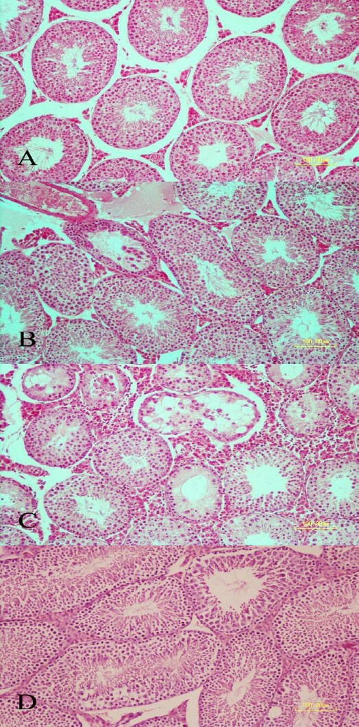
Figure 3.

Testicular sections stained with hematoxylin and eosin (bar = 100 µm). A: Control group section showing normal seminiferous tubules morphology. B: A few seminiferous tubules revealing mild degenerative changes on day 3 after LPS administration. C: Photomicrograph showing seminiferous tubules at days 30 following LPS administration. Severe degenerative changes are observed. Depletion and vacuolation of epithelium with decreased number of germinal layers are seen in some of the seminiferous tubules. D: Photomicrograph showing seminiferous tubules at day 60 of LPS administration. Normal histomorphology of tubules is preserved
