
Figure 5.
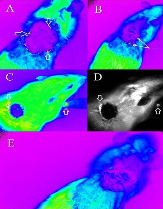
Tracking of the cGFP-expressing mesenchymal stem cells (MSCs) using the Kodak in vivo Imaging System F-Pro device. The MSCs were tracked in treated rats in color mode (A) 1 week after treatment (B) 2 weeks after treatment (C) 3 weeks after treatment. (D) Indicates the cells shown in (C) in gray mode. (E) The control rat treated with gauze vaseline after 1 week. The cGFP expressing MSCs could be detected in close proximity to the burnt area as evident by their greater intensity. The cells are indicated by the arrows.