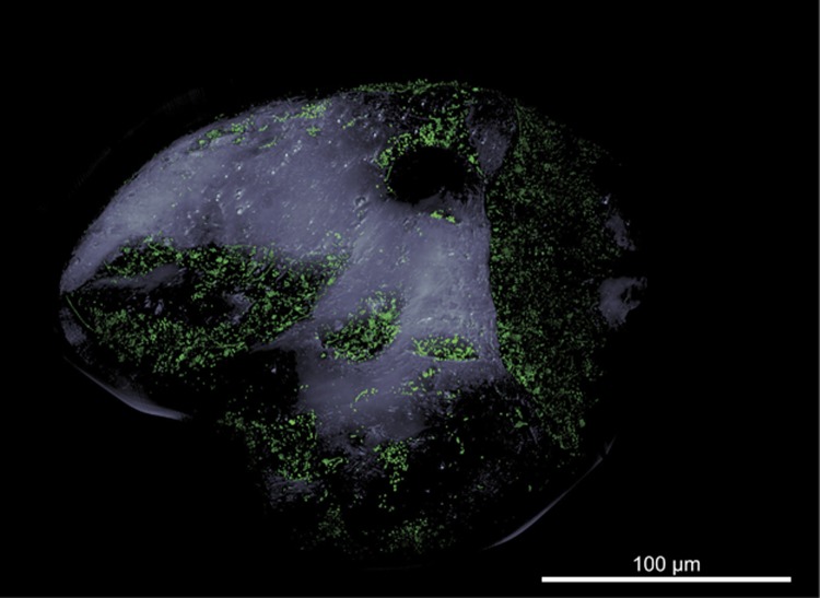
Figure 1.
Microbial colonization of a sand grain. Confocal laser scanning micrograph showing SYBR green I-stained microbial cells on a sand grain visualized as three-dimensional reconstruction. The grain’s surface was visualized by transmitted light microscopy. Note the bare surfaces of convex and exposed areas in contrast to protected areas dominated by macrotopography, which are densely populated by microbes.

