Figure 5.
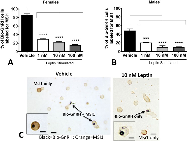
Leptin stimulation of MSI1 in gonadotropes. Monolayer cultures of pituitary cells were stimulated with leptin for 3 hours and then exposed to biotinylated GnRH for the last 10 minutes of culture. After fixation, cells were dual labeled for bio-GnRH and MSI1. MSI proteins are found (A) in 84% of bio-GnRH bound gonadotropes in females and (B) in ∼50% of bio-GnRH cells in males. After 3 hours of leptin stimulation, there is a dramatic loss in expression of MSI1 proteins in both males and females. ANOVA followed by Bonferroni multiple comparisons test. (C) A sample photomicrograph demonstrates the dual-labeled cells containing black label for bio-GnRH and orange label for MSI1 (left). A field treated with 10 nM leptin and the loss of orange labeling for MSI1 in the biotinylated GnRH-labeled cells (right). The label for bio-GnRH is stronger, as would be expected following leptin stimulation. Low magnification bar = 20 μm; high magnification bars = 10 μm. *P < 0.05; **P < 0.005; ***P < 0.0005; ****P < 0.0001.

