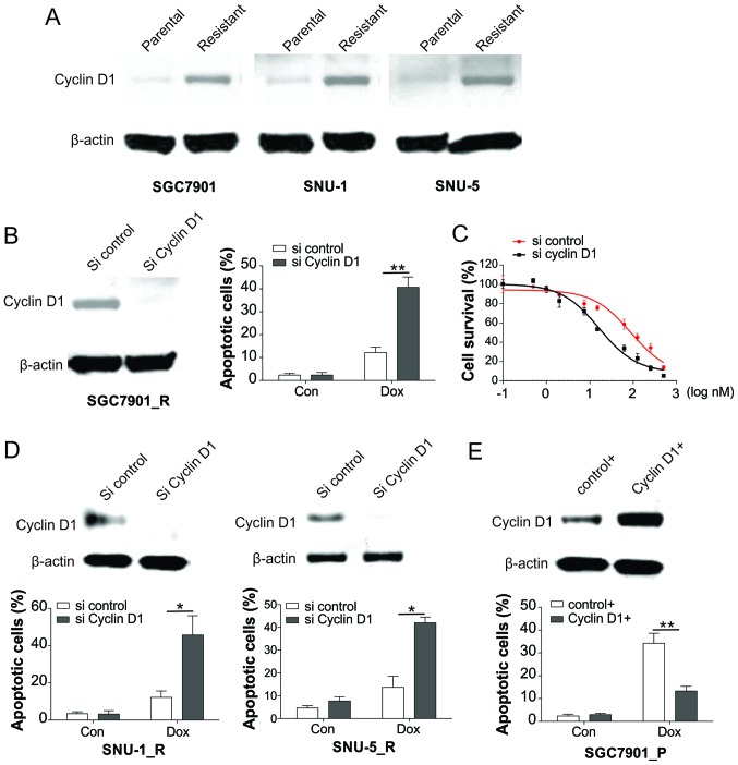
Figure 2.
Induction of cyclin D1 results in Dox resistance in GC cells. (A) Expression of cyclin D1 in SGC7901, SNU-1 and SNU-5 parental and resistant cells was analyzed by western blot analysis. (B) SGC7901 resistant cells were transfected with cyclin D1 siRNA, and then the expression of cyclin D1 was analyzed by western blot analysis (left), while the apoptotic cells following Dox treatment were analyzed by Hoechst 33258 staining (right). (C) SGC7901 resistant cells underwent transfection with cyclin D1 siRNA, followed by treatment with Dox, and then the viability of cells was examined using Cell Counting kit-8 assay. (D) SNU-1 and SNU-5 resistant cells were transfected with cyclin D1 siRNA. The expression of cyclin D1 was analyzed by western blot assay (upper), and the apoptotic cells following Dox treatment were analyzed by Hoechst 33258 staining (lower). (E) SGC7901 parental cells were transfected with cyclin D1 plasmid, and the expression of cyclin D1 was analyzed by western blot assay (upper), while the apoptotic cells after Dox treatment were analyzed by Hoechst 33258 staining (lower). Error bars represent the standard deviation of triplicate detection. *P<0.05 and **P<0.01. GC, gastric cancer; Dox, doxorubicin; siRNA, small interfering RNA; Con, control.

