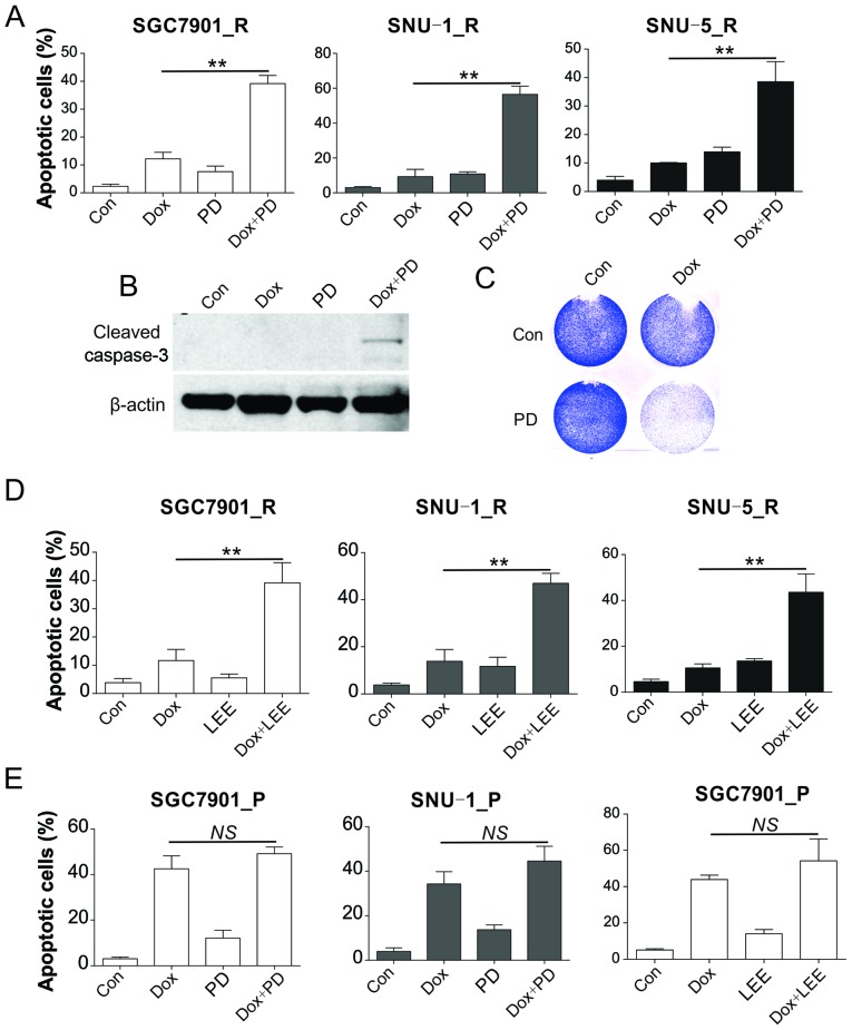
Figure 3.
CDK inhibitors sensitized the resistant GC cells to Dox. (A) SGC7901_R, SNU-1_R and SNU-5_R cells were treated with 20 nM Dox and/or 40 nM PD-0332991 for 24 h. The apoptotic cells were analyzed by Hoechst 33258 staining. (B) Caspase-3 expression was analyzed by western blot analysis, and (C) cell viability was examined by crystal violet staining in SGC7901_R cells treated with 20 nM Dox and/or 40 nM PD-0332991 for 24 h. (D) Apoptosis was analyzed by Hoechst staining in SGC7901_R, SNU-1_R, and SNU-5_R cells treated with 20 nM Dox and/or 20 nM LEE011 for 24 h. (E) Apoptosis was analyzed by Hoechst staining in SGC7901_P and SNU-1_P cells treated with 20 nM Dox and/or 40 nM PD-0332991 or 20 nM LEE011 for 24 h. Error bars indicate the standard deviation of triplicate detection. **P<0.01. NS, non-significant; GC, gastric cancer; Dox, doxorubicin; _P, parental; _R, resistant; Con, control.

