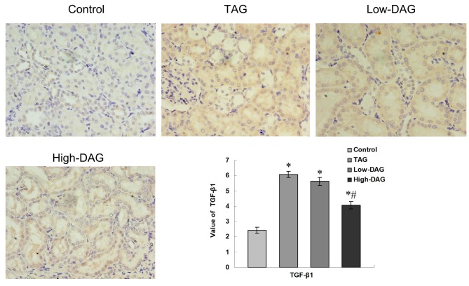
Figure 5.
Immunohistochemical staining of TGF-β1 in renal tissues from experimental rats (magnification, ×400). Quantification of the value of the TGF-β1-stained area was analyzed using a secondary scoring method. Data are represented as the mean ± standard deviation (n=10). *P<0.05 vs. control group; #P<0.05 vs. TAG group. DAG, diacylglycerol; TAG, triacylglycerol; TGF-β1, transforming growth factor-β1.

