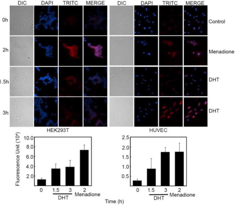
Figure 4.
Detection of ROS in HEK293T and HUVEC cells. Cells in culture were either treated with DHT (10 µM) for 1.5 and 3 h (HUVEC 0hr vs 3hr *P=0.0039, 0hr vs menadione *P=0.029 and HEK293T 0hr vs menadione *P=0.020) or menadione (200 µM) for 2 h. Presence of ROS was detected using the deep red CellRox reagent having tetramethylrhodamine (TRITC) as the fluorescent dye. Experiments are done in triplicates with error bars representing ±SEM. A) Microscopic images of cells showing formation of ROS B) Quantitation of ROS measured as the fluorescence intensity within the cell.

