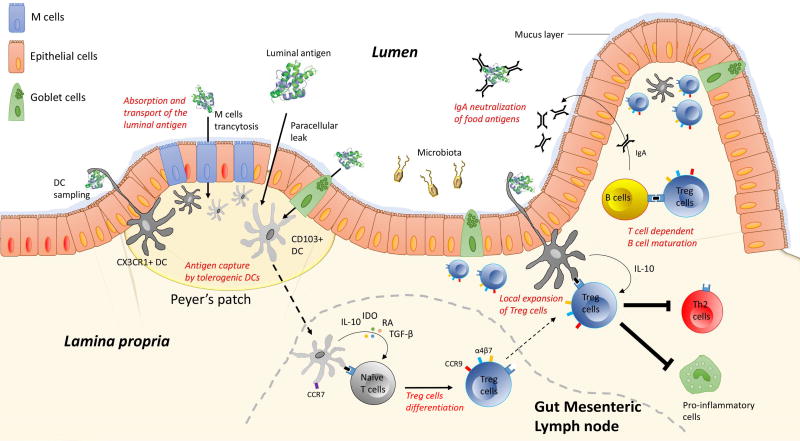
Figure 1.
Schematic overview of the stepwise mechanisms involved in the development and maintenance of oral tolerance. Upon ingestion and digestive process, luminal antigen are absorbed and transported through intestinal epithelial cells thus enabling antigenic presentation to underlying immune cells. This complex process might include: 1) paracellular diffusion, 2) endocytosis via microfold cells and globlet cells, and 3) trans-epithelial DC sampling of the luminal antigen. In specific areas of the epithelium, called Peyer’s patches, luminal antigens are taken up by CD11c+CD103+DCs or CD11b+CX3CR1+DCs. While CX3CR1+DCs remain located in the lamina propria, antigen-loaded CD103+ DCs migrate to the mLN in a CCR7-dependent mechanism to promote the differentiation of naïve T cells into regulatory T (Treg) cells. After their generation in lymph nodes, Tregs up-regulate CCR9 and integrin α4β7 to home back to the lamina propria where they undergo local expansion to induce oral tolerance. In this process Treg cells play a major role in: 1) promoting B-cell class switching to produce the non-inflammatory IgA responses, 2) inducing T-cell anergy of effector cells, and 3) inhibiting downstream pro-inflammatory cells.

