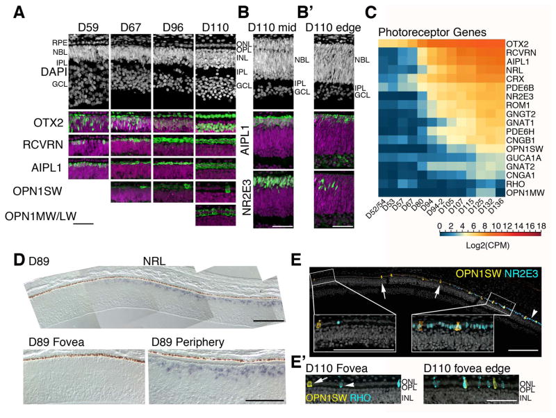
Figure 4. Photoreceptor development in the human fetal retina.
A. Human fetal retinae from ages D59–D110 were stained with photoreceptor markers. Each of the photoreceptor markers are shown in green and nuclei were counterstained with DAPI (magenta). A single layer of photoreceptors was already present at D59, some of which were AIPL1+ and RCVRN+. The first OPN1SW+ cell was detected at D67 and OPN1MW/LW was detected at D110. Foveas older than D67 were generally defined as a zone free of S-Opsin and NR2E3 immunoreactivity. B. Retinal structure and AIPL1 and NR2E3 expression were compared between mid-temporal (B) and temporal peripheral edge (B′) of a D110 retina. Nuclear layers were not separated yet and NR2E3 expression was just beginning at the retinal edge. C. Heatmap for RNA-seq analysis of pan photoreceptor, cone, and rod genes show a steady increase over time. Blue indicates low and red indicates high gene expression. D. In situ hybridization of NRL mRNA at D89. The composite image shows the NRL-free fovea; NRL expression rapidly increases from the foveal edge and reaches the periphery (bottom right panel). E. A composite image of D110 fovea and 450 μm perifovea towards temporal retina. Immunostaining with OPN1SW (yellow) and a rod-specific marker NR2E3 (blue) showed a sharp increase in rod generation adjacent to the fovea. E′. A subset of these rods expressed RHO (blue). Scale bars for A–C: 50 μm.

