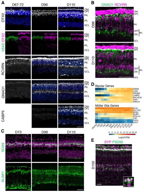
Figure 5. Later cell types are generated by D73 in the fovea and bipolar cell process stratification is observed by D96.
A. Fovea from D67–110 retinae were immunostained with OTX2 (first row: white, photoreceptors, bipolars); VSX2 and OTX2 (green and magenta, respectively; bipolar cells); RCVRN (white, OFF bipolar cells); GNAO1 (white, ON bipolar cells); and CABP5 (white, cone bipolar cells). Arrow heads point to VSX2+OTX2+ double-labeled bipolar cells. GNAO1 was first observed at D96 and CABP5 at D110. B. Higher magnification of D96 and D110 fovea double-labeled with GNAO1 and RCVRN show axonal arborizations in the IPL that were stratified into ON and OFF sublaminae by D96. C. Sections of D73–110 foveas were immunostained with SOX9 and RLBP1 (both green) revealed that a row of Müller glia was present in the fovea at D73 and RLBP1 expression got more elaborated over time. D. Log2 transformed Counts Per Million (CPM) values of select bipolar and Müller glia genes show a steady increase in gene expression beginning at D67–D80. Expression values plotted from high to low based on expression at D136. Blue to red indicates low to high gene expression. E. Immunostaining of the fovea of a D150 retina. In the IPL, SYP and PSD95 were expressed opposed to each other indicative of putative synapses. Scale bars: 50 μm. See also Figure S3.

