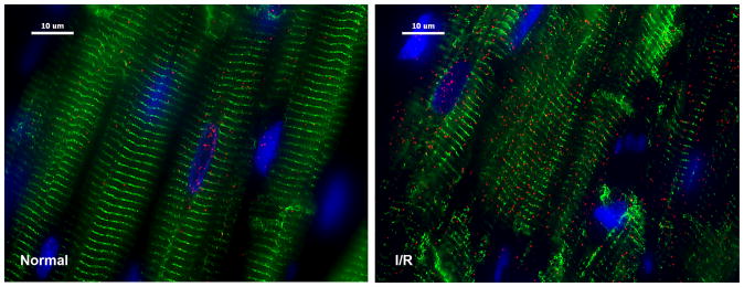
Figure 5. Chymase inside dog cardiomyocytes during ischemia/reperfusion.
Adult dogs subjected to 60 minutes of LAD occlusion and 100 minutes of reperfusion (right panel) or normal controls (left panel). Ischemia/reperfusion LV led to a marked increase in chymase (red) in cardiomyocytes with breakdown of desmin (green, right). I/R: ischemia/reperfusion; blue: DAPI.158 Reproduced with permission of Plos One.

