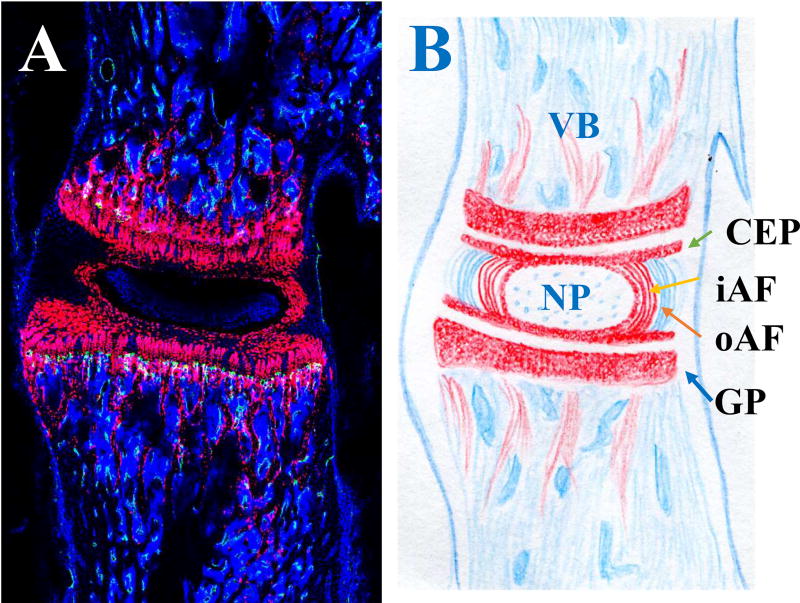
Figure 1. Mouse lumbar intervertebral disc (IVD).
A. Sagittal section of a Col2CreER;R26- tdTomato mouse IVD. B: schematic drawing of the vertebral body (VB)-IVD-VB motion segment. Red: type II collagen expressing cells; Blue: cell nuclei stained with DAPI. NP: nucleus pulposus; CEP: cartilaginous endplate; iAF: inner annulus fibrosus (AF); oAF: outer AF; GP: growth plate.

