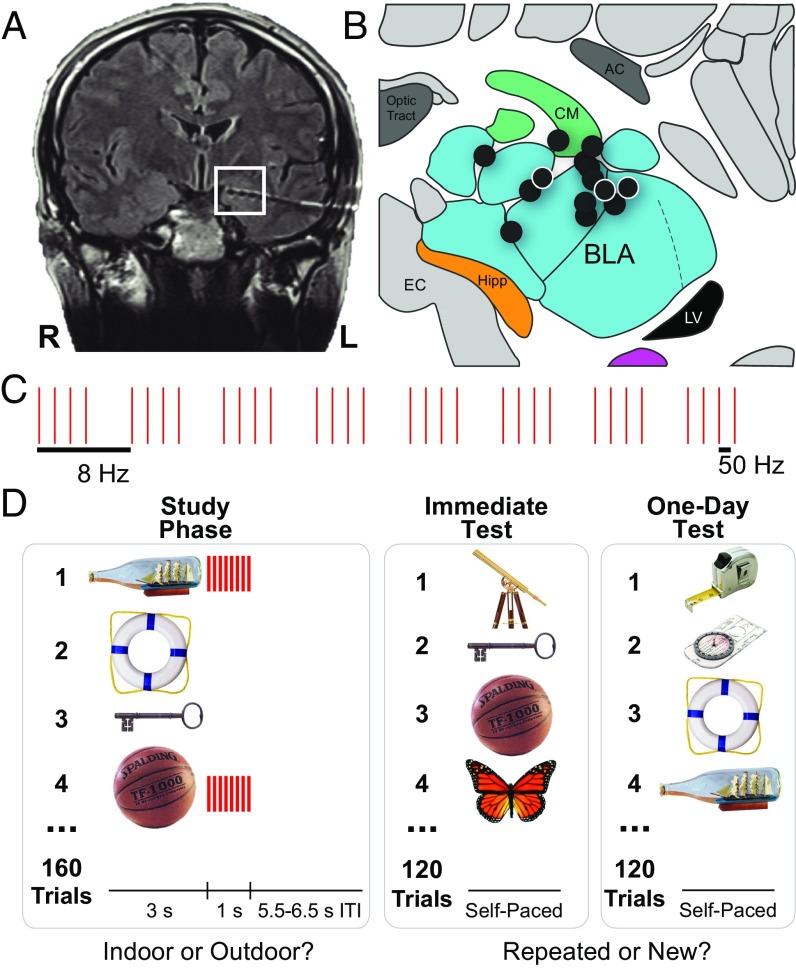
Fig. 1.
The procedure used to stimulate the human amygdala and to test recognition memory. (A) A representative postoperative coronal MRI showing electrode contacts in the amygdala (white square). (B) Illustration of left amygdala (coronal slice) with black circles indicating estimated centroids of bipolar stimulation in or near the BLA in all 14 patients. (See Fig. S1 for more precise localizations.) White borders denote right-sided stimulation. All patients had at least one bipolar stimulation contact in the BLA. AC, anterior commissure; CM, centromedial complex of the amygdala; EC, entorhinal cortex; Hipp, head of the hippocampus; LV, lateral ventricle (temporal horn). Adapted with permission from ref. 54, copyright Elsevier 2007. (C) Schematic of the 1-s stimulation pulse sequence (each pulse = 500 μs biphasic square wave; pulse frequency = 50 Hz; train frequency = 8 Hz). (D) Schematic of the recognition-memory task in which the amygdala was stimulated after the presentation of half of the objects in the study phase and recognition memory was tested on unique subsets of images immediately and one day after the study phase.

