Fig. 1.
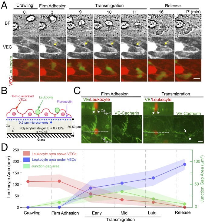
Three-dimensional endothelial deformation is associated with the remodeling of VEC junctional gaps during leukocyte paracellular diapedesis. (A) Epifluorescent live-cell imaging of VEC junctions stained by the CY5 cell membrane dye and a leukocyte stained by the FITC dye. The yellow arrow indicates the location where an intercellular gap is formed during diapedesis. Merged-color images show the diapedesis stages: crawling, firm adhesion, transmigration, and final release. (Scale bar, 10 μm.) (B) The experimental setup for 3DTFM during leukocyte diapedesis. (C) Three-dimensional confocal images of leukocytes fixed during diapedesis as described in Materials and Methods. Orthogonal (XZ) projection demonstrates a leukocyte is crossing the VEC border. White arrows indicate that a VE-Cadherin gap associated with the extravasating leukocyte. (Scale bar, 10 μm.) (D) Time evolution of the area projection (XY plane) of a leukocyte and VEC junctional gap during diapedesis. Data are representative of three independent experiments with two to three fields in each experiment group. Data are expressed as mean ± SEM.

