Figure 1.

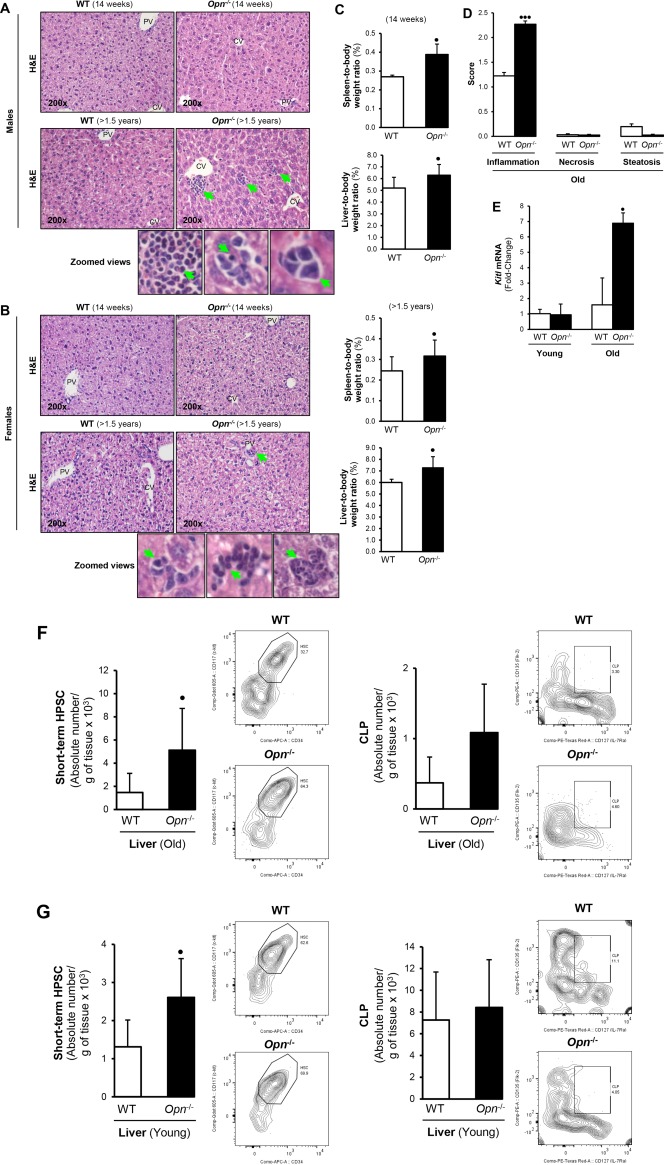
Opn −/− mice show increased hepatic hematopoietic progenitors compared to WT mice. (A,B) H&E staining reveals small aggregates of cells with intensely basophilic nuclei and clusters of cells in the liver of (top panels) young (14‐16 weeks;) and (bottom panels) old (>1.5 years; green arrows in panels and zoomed views) Opn −/− mice. (C) Spleen‐to‐body weight and liver‐to‐body weight ratios in young males and females show hepatosplenomegaly in Opn −/− compared to WT mice. (D) Pathology scores from old Opn −/− and WT mice. (E) Kitl mRNA in liver of young and old Opn −/− and WT male mice. (F) Opn deletion increases CD34+ and CD127+ cell hepatic population in old and young mice. (G) Old and young Opn −/− mice displayed increased Lin– Sca‐1+ c‐kit+ CD34+ CD135+ short‐term HPSCs and Lin– Sca‐1+ c‐kit+ CD127+ CD135+ CLPs compared to WT littermates. Bar graphs and representative flow plots are shown. Liver leukocytes were isolated and gated using SSC/FSC, viability dye (to exclude dead cells), single‐cell population (to exclude doublets), and Lin– Sca‐1+ c‐kit+ (to identify hematopoietic cells). Results are expressed as mean ± SD; n = 6/group, • P < 0.05, •• P < 0.01, and ••• P < 0.001 for Opn −/− versus WT mice (both genders). Abbreviations: APC, antigen‐presenting cell; CV, central vein; FSC, forward scatter; H&E, hematoxylin and eosin; Lin– Sca‐1+ c‐kit+, lineage‐negative, stem cell antigen‐1‐positive, and c‐kit receptor‐positive; PE, phycoerythrin; PV, portal vein; SSC, side scatter.
