
Figure 2.
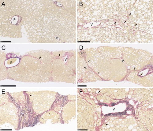
Histologic localization of collagen and elastin fibers in liver biopsy tissues from patients with NAFLD. Collagen and elastin fibers were stained red and black, respectively, by EVG staining. Fibrotic areas in liver biopsy specimens were mainly composed of collagen fibers. (A) Native collagen fibers were found in portal tracts and hepatic veins. (B) Zone 3 perisinusoidal fibrosis (arrowheads) was a characteristic finding of stage 1 NAFLD and was also seen in stages 2‐4. (C) Patients with zone 3 and portal fibrosis were categorized as stage 2. (D,E) Bridging fibrosis (arrows) that links vascular structures is a hallmark of advanced NAFLD (stages 3/4). Thick and extensive bridging fibrosis was infrequent in stage 3 but was common in stage 4. (F) Increased elastin deposition was often found in areas of thick bridging fibrosis and thickened venous walls but was uncommon in areas of perisinusoidal fibrosis. Stage 0 (A), 1 (B), 2 (C), 3 (D), and 4 (E); EVG stain (A‐F). Scale bars, 500 μm (C), 250 μm, (A,D,E), 100 μm (B,F). Abbreviations: P, portal tract; V, hepatic vein.