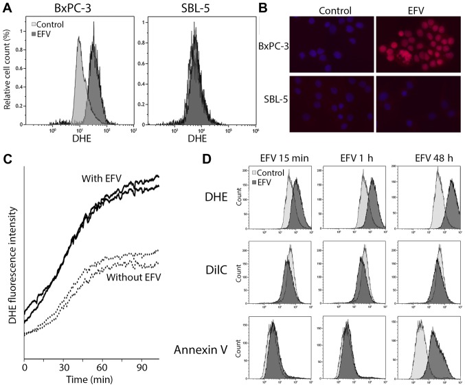
Figure 3.
EFV-induced oxidative stress. (A) Flow cytometric assessment and (B) immunostaining of oxidative stress induction with DHE in BxPC-3 pancreatic cancer cells and SBL-5 primary fibroblasts treated with EFV (40 µmol/l for 1 h). (C) Time kinetics of oxidative stress induction after EFV treatment (40 µmol/l) in life cell imaging in BxPC-3 cells. Fluorescence intensity was scanned at two positions with EFV (solid lines) and two positions without EFV (dotted lines). (D) Simultaneous flow cytometric assessment of oxidative stress (DHE), mitochondrial membrane potential (DilC) and apoptosis (Annexin V) after EFV treatment. DHE, Dihydroethidium; EFV, Efavirenz.

